Skip to content
 
 
Skip to product information
1 of 12

Revo-NX130

Revo-NX130

Excellent

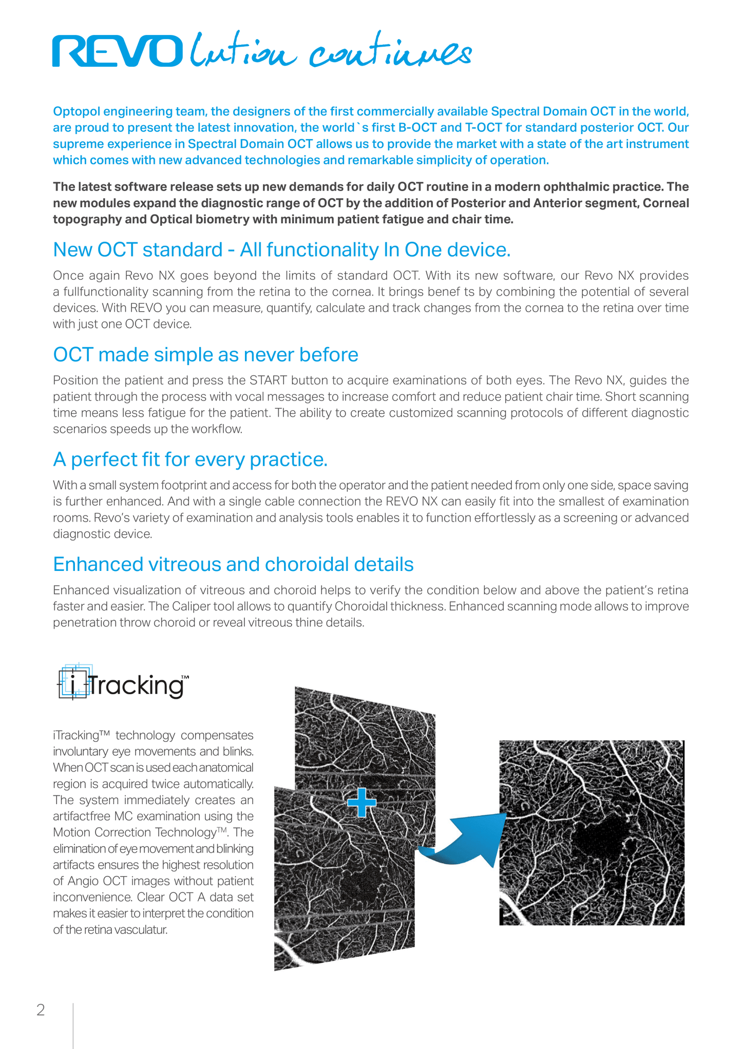
Optopol engineering team, the designers of the first commercially available Spectral Domain OCT in the world,
are proud to present the latest innovation, the world`s first B-OCT and T-OCT for standard posterior OCT. Our
supreme experience in Spectral Domain OCT allows us to provide the market with a state of the art instrument
which comes with new advanced technologies and remarkable simplicity of operation.
The latest software release sets up new demands for daily OCT routine in a modern ophthalmic practice. The
new modules expand the diagnostic range of OCT by the addition of Posterior and Anterior segment, Corneal
topography and Optical biometry with minimum patient fatigue and chair time.
New OCT standard - All functionality In One device.
Once again Revo NX goes beyond the limits of standard OCT. With its new software, our Revo NX provides
a fullfunctionality scanning from the retina to the cornea. It brings benef ts by combining the potential of several
devices. With REVO you can measure, quantify, calculate and track changes from the cornea to the retina over time
with just one OCT device.
OCT made simple as never before
Position the patient and press the START button to acquire examinations of both eyes. The Revo NX, guides the
patient through the process with vocal messages to increase comfort and reduce patient chair time. Short scanning
time means less fatigue for the patient. The ability to create customized scanning protocols of different diagnostic
scenarios speeds up the workflow.
A perfect fit for every practice.
With a small system footprint and access for both the operator and the patient needed from only one side, space saving
is further enhanced. And with a single cable connection the REVO NX can easily fit into the smallest of examination
rooms. Revo’s variety of examination and analysis tools enables it to function effortlessly as a screening or advanced
diagnostic device.
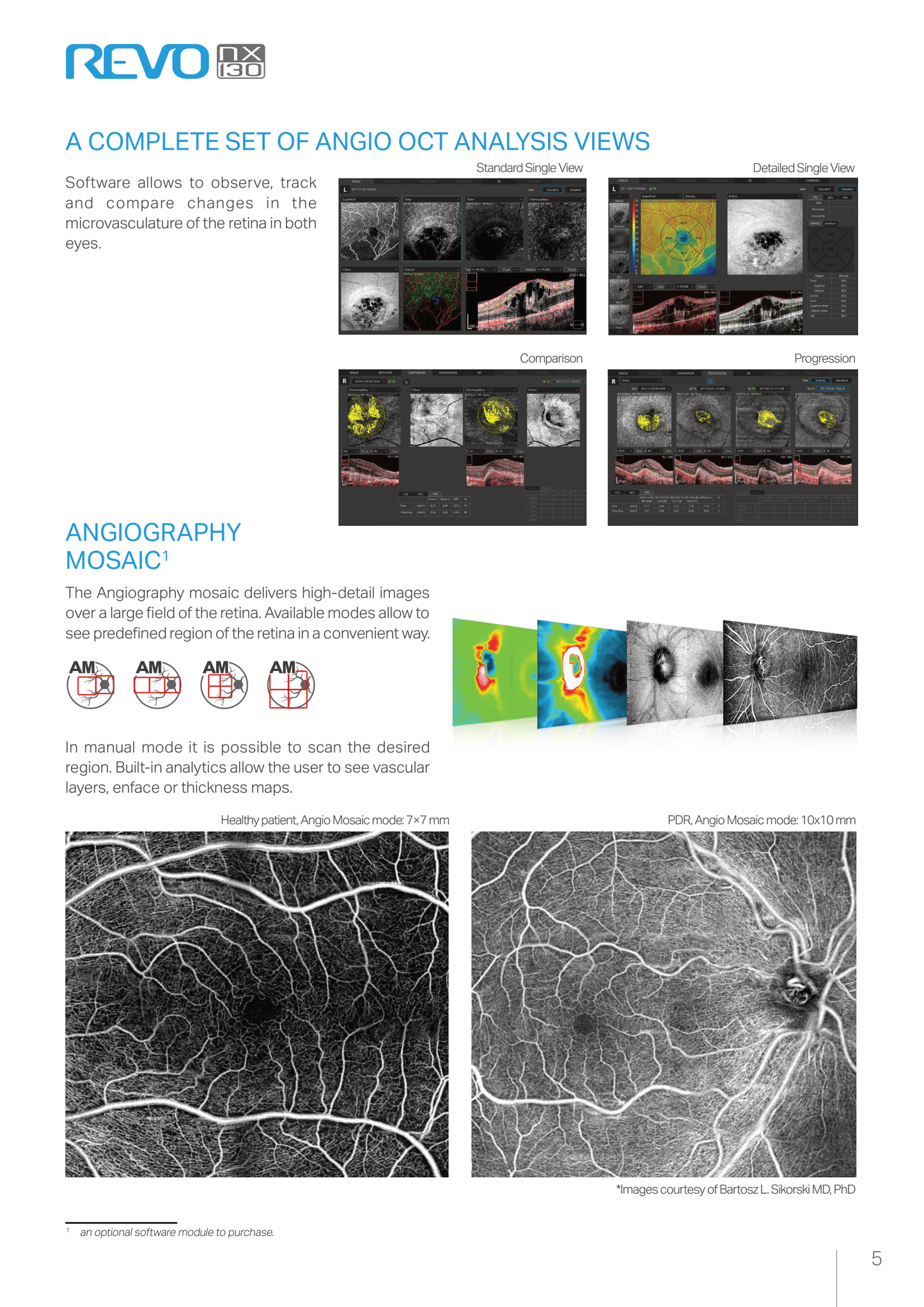
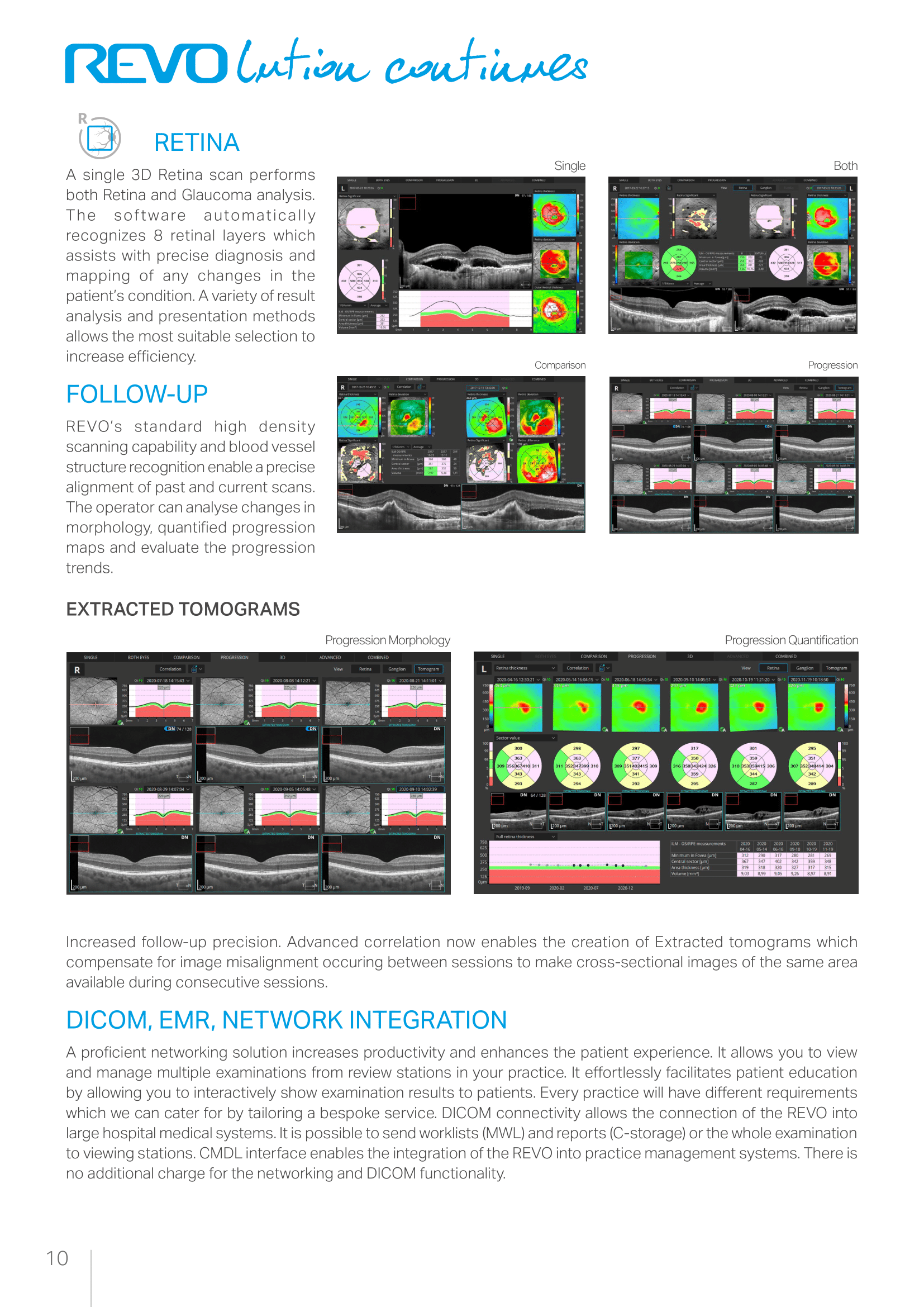
Enhanced vitreous and choroidal details
Enhanced visualization of vitreous and choroid helps to verify the condition below and above the patient’s retina
faster and easier. The Caliper tool allows to quantify Choroidal thickness. Enhanced scanning mode allows to improve
penetration throw choroid or reveal vitreous thine details.

  • Easy Setup
  • 24/7 VIP Support
View full details

Key Specifications

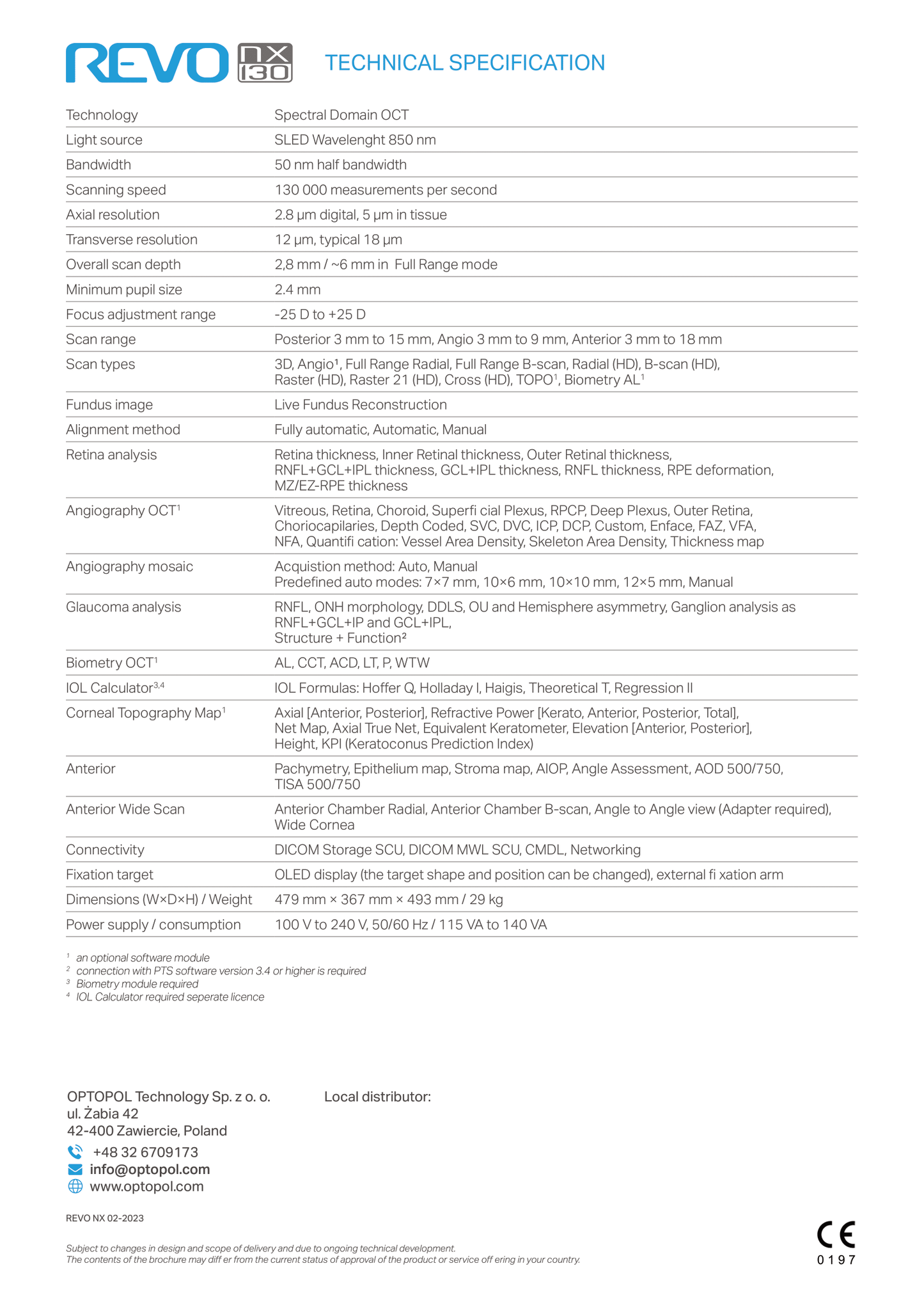
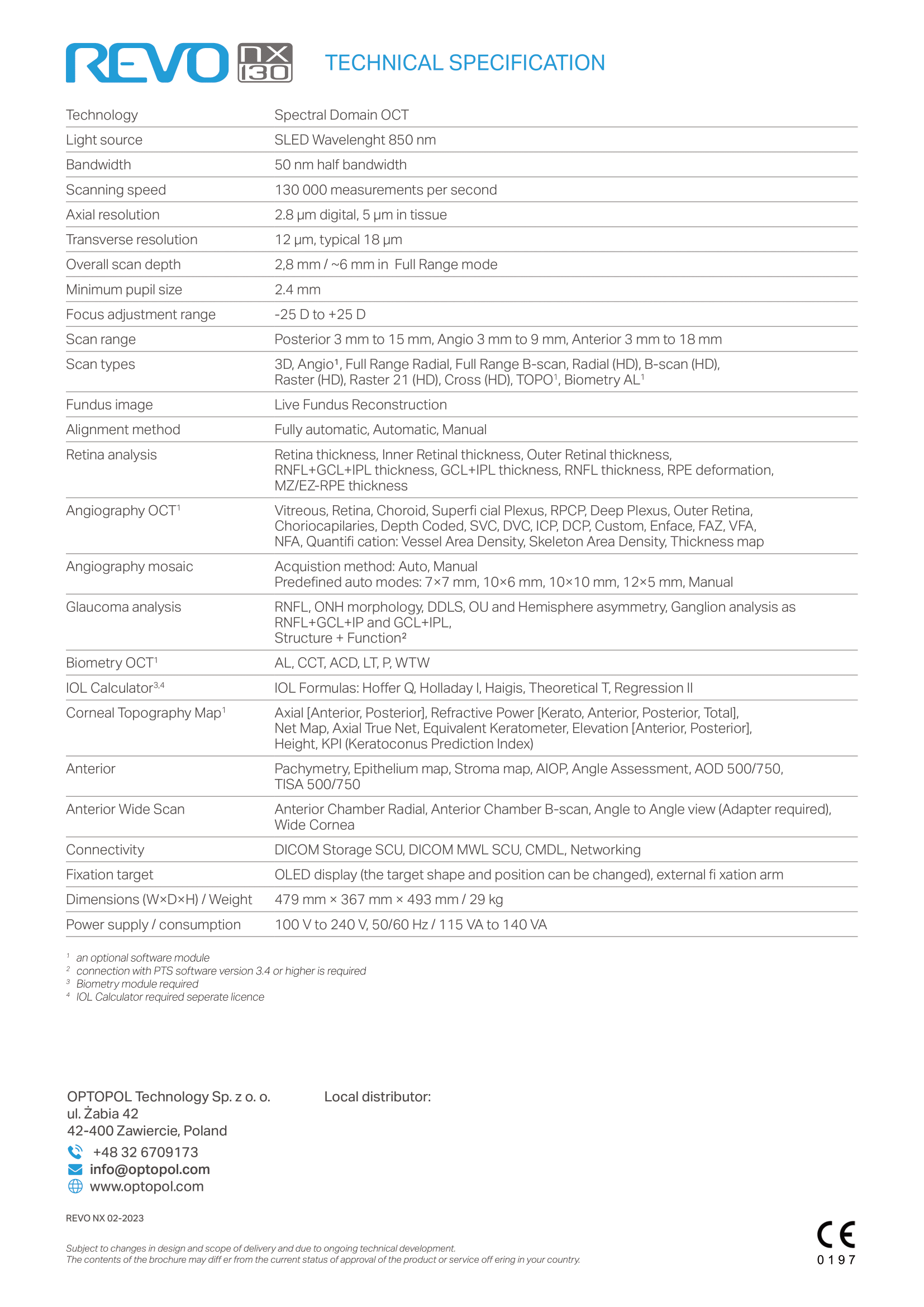
Scanning Speed

130,000 A-scans per second

Axial Resolution

2.8 µm digital, 5 µm in tissue

Transverse Resolution

12 µm, typical 18 µm

Overall Scan Depth

2.8 mm; approximately 6 mm in Full Range™ mode

Pupil Size

Minimum 1.7 mm

Focus Adjustment Range

-25D to +25D

Light Source

Superluminescent Diode (SLED), 830 nm wavelength, 50 nm half bandwidth

Dimensions (L×W×H)

479 mm × 367 mm × 493 mm

Weight

29 kg

Power Supply

100 V to 240 V, 50/60 Hz, 90 VA to 110 VA

Fixation Target

OLED display with customizable target shape and position; external fixation arm

Connectivity

DICOM Storage SCU, DICOM MWL SCU, CMDL, Networking

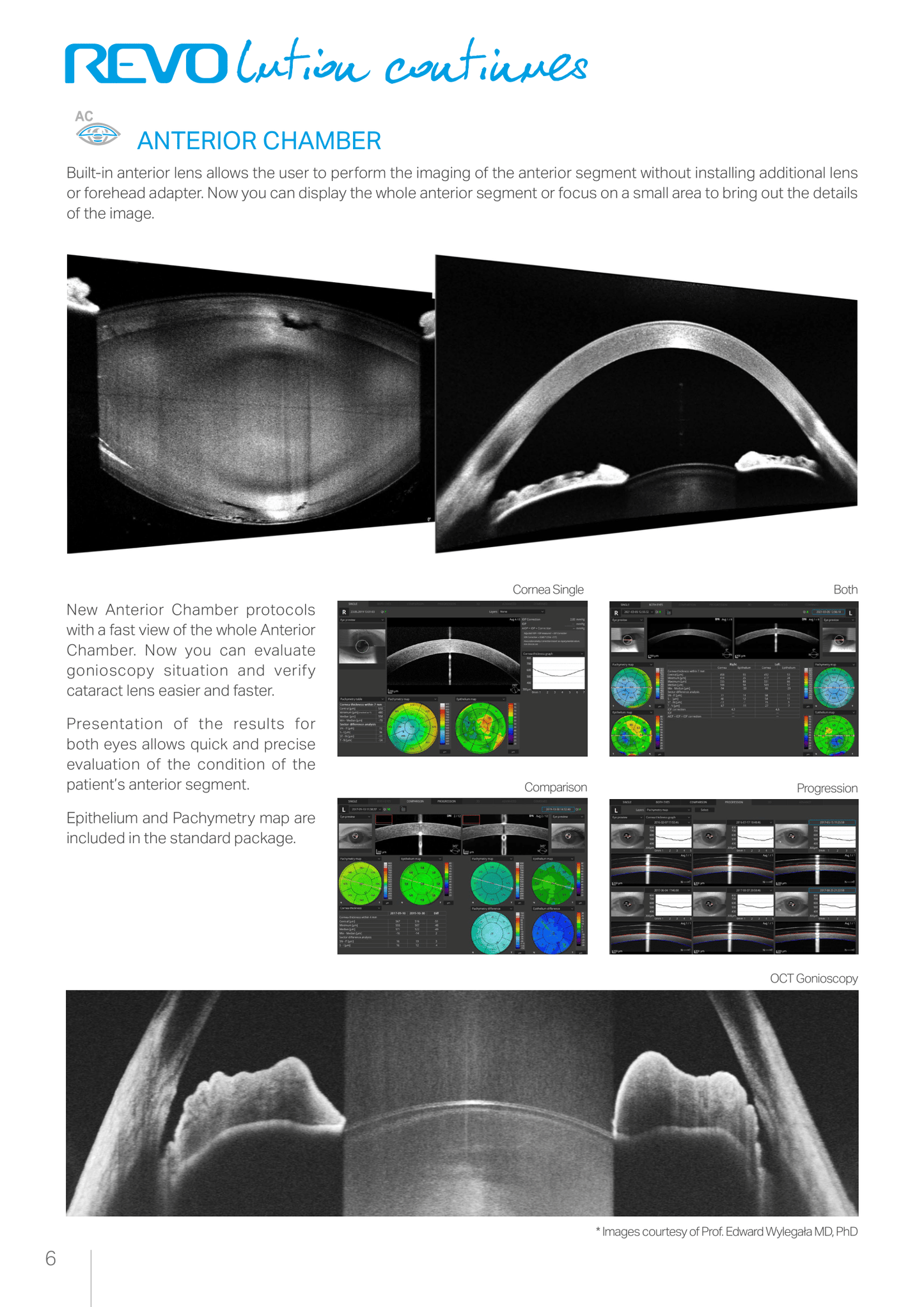
Anterior Chamber Imaging & Analysis

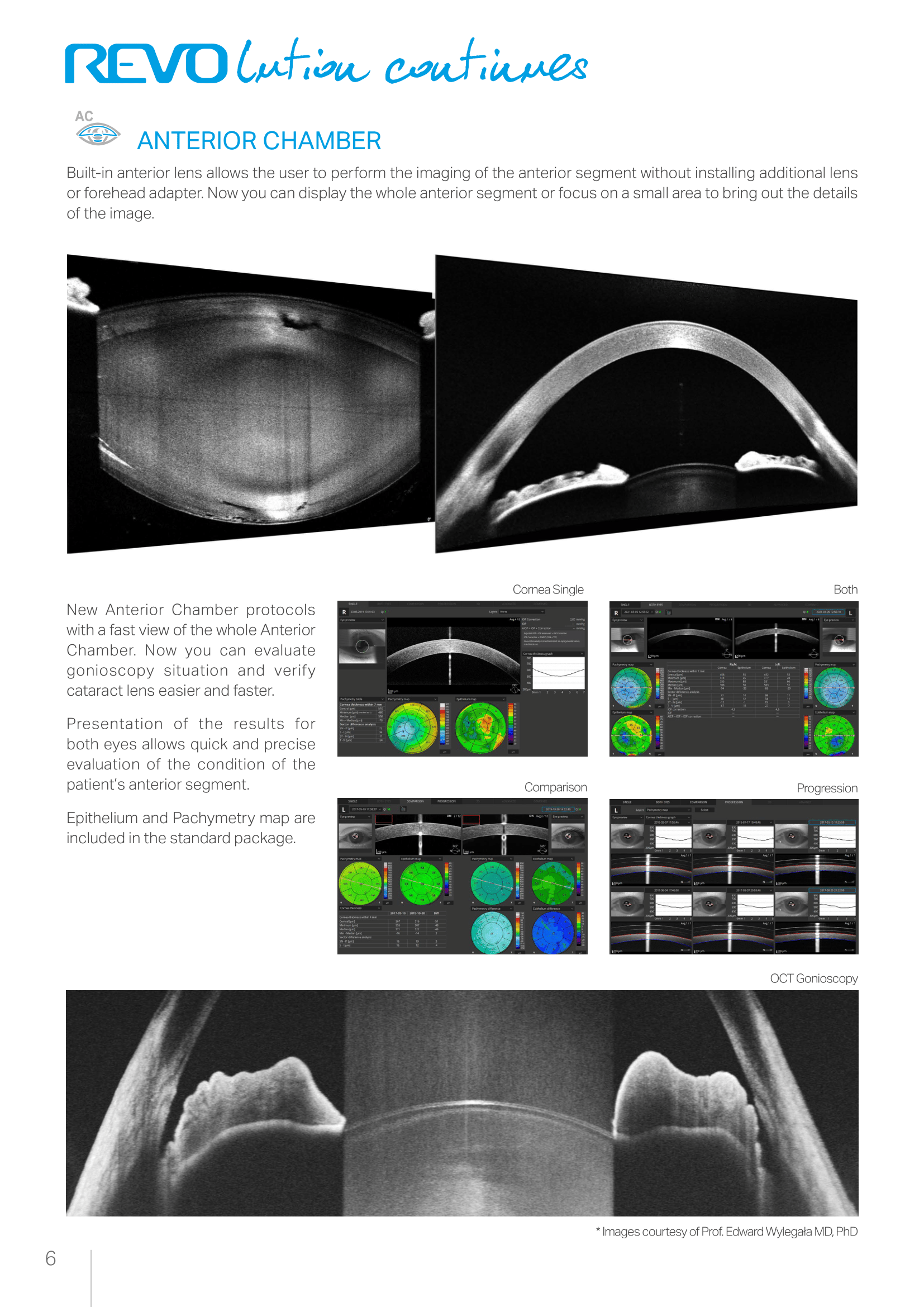
The REVO NX 130 offers advanced anterior segment imaging, delivering precise and high-resolution scans of the anterior chamber, cornea, iris, and lens. Its imaging technology provides detailed cross-sectional views that are essential for assessing conditions like glaucoma, cataracts, and corneal abnormalities.

With a wide focus adjustment range (-25D to +25D) and minimum pupil size of 1.7 mm, it captures sharp images even in challenging clinical scenarios. The device’s 3D imaging and B-scan modes enable comprehensive visualization of the anterior chamber depth and angle structures, supporting accurate diagnosis and treatment planning.

Clinicians can rely on the REVO NX 130 for consistent, reproducible measurements critical for preoperative assessments, surgical planning, and disease monitoring within the anterior segment.

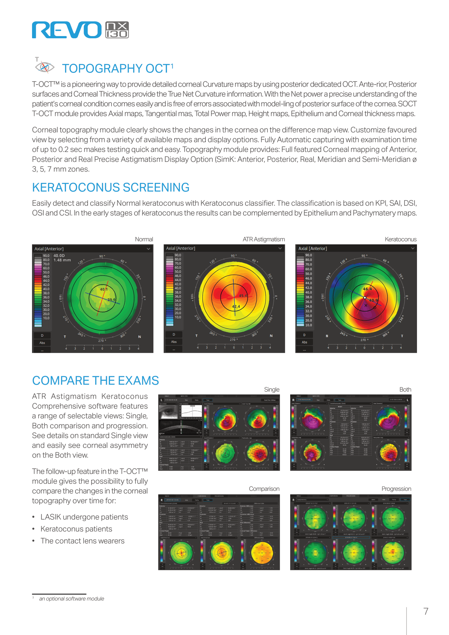
Keratoconus Screening & Topography OCT Features

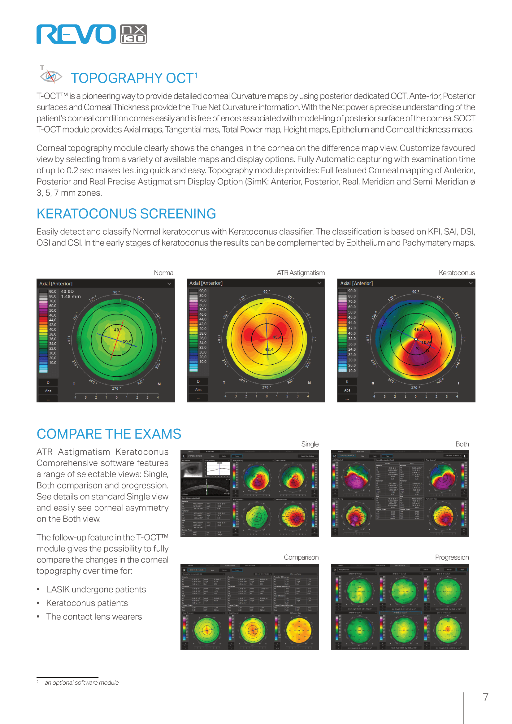
The REVO NX 130 is equipped with advanced Topography OCT capabilities that provide detailed mapping of the corneal surface and thickness, making it a powerful tool for keratoconus screening and early detection of corneal ectasia. Utilizing high-resolution OCT imaging combined with precise corneal topography, it enables clinicians to identify subtle changes in corneal curvature and thickness before clinical symptoms appear.

Its 3D imaging and depth-resolved tomography allow for comprehensive analysis of both the anterior and posterior corneal surfaces, delivering accurate pachymetry maps and elevation data. This detailed information supports the differentiation between normal corneas and those affected by keratoconus or other corneal pathologies, enabling timely intervention.

The device’s software integrates keratoconus screening algorithms that automatically flag suspicious cases based on topographic indices, improving diagnostic confidence and reducing the risk of missed diagnoses. Additionally, the OCT-Angiography (OCT-A) module complements these features by visualizing corneal vascular changes that may be associated with disease progression.

With its fast scan speed, user-friendly interface, and comprehensive topography reports, the REVO NX 130 stands out as a cutting-edge solution for clinicians focusing on corneal health and refractive surgery planning.