Skip to content
 
 
Skip to product information
1 of 9

Revo-HR

Revo-HR

Excellent

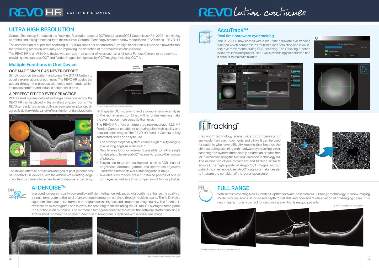
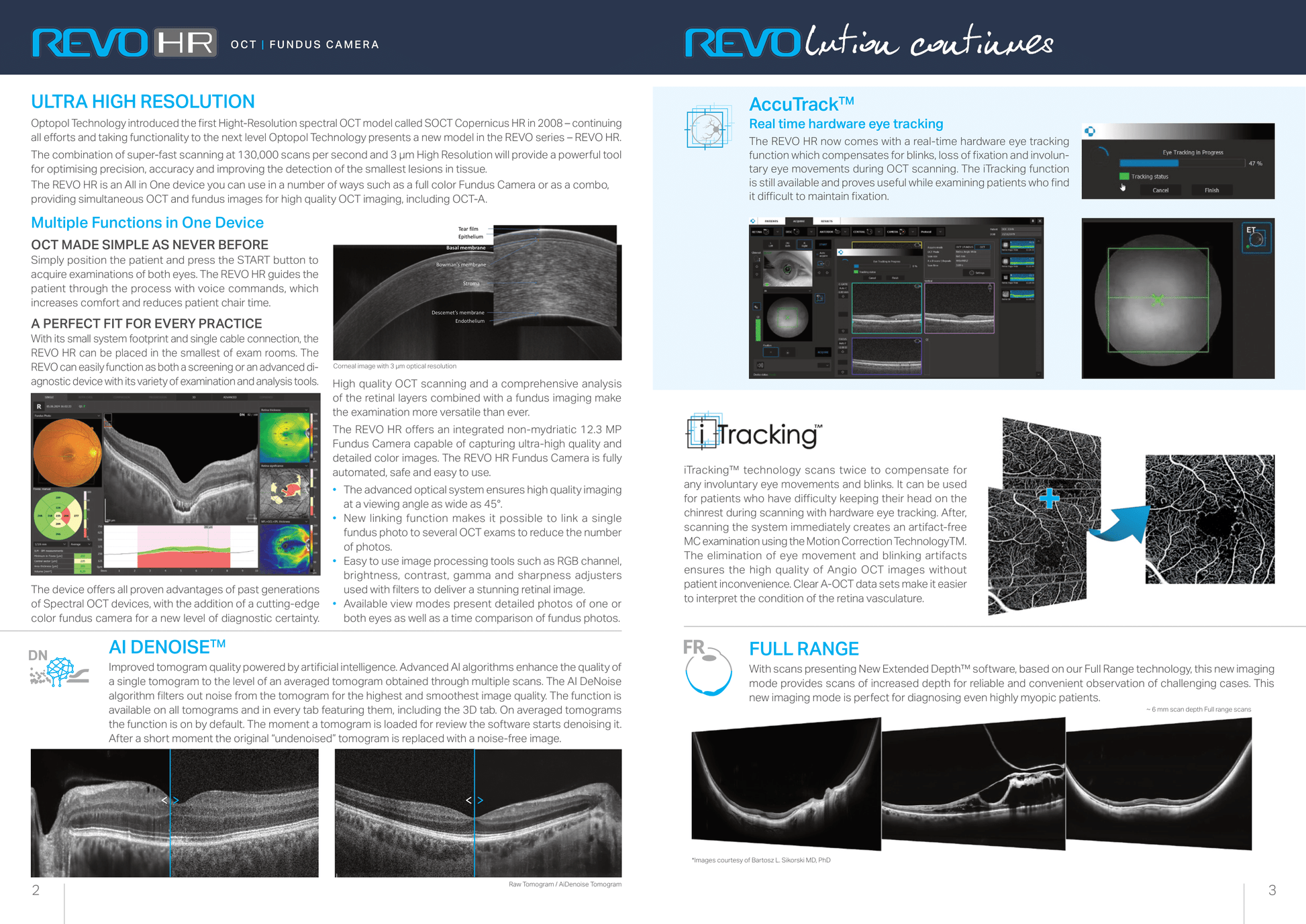
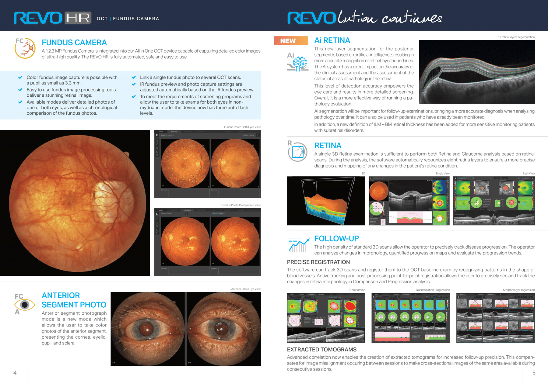
Optopol Technology introduced the first Hight-Resolution spectral OCT model called SOCT Copernicus HR in 2008 – continuing
all efforts and taking functionality to the next level Optopol Technology presents a new model in the REVO series – REVO HR.
The combination of super-fast scanning at 130,000 scans per second and 3 μm High Resolution will provide a powerful tool
for optimising precision, accuracy and improving the detection of the smallest lesions in tissue.
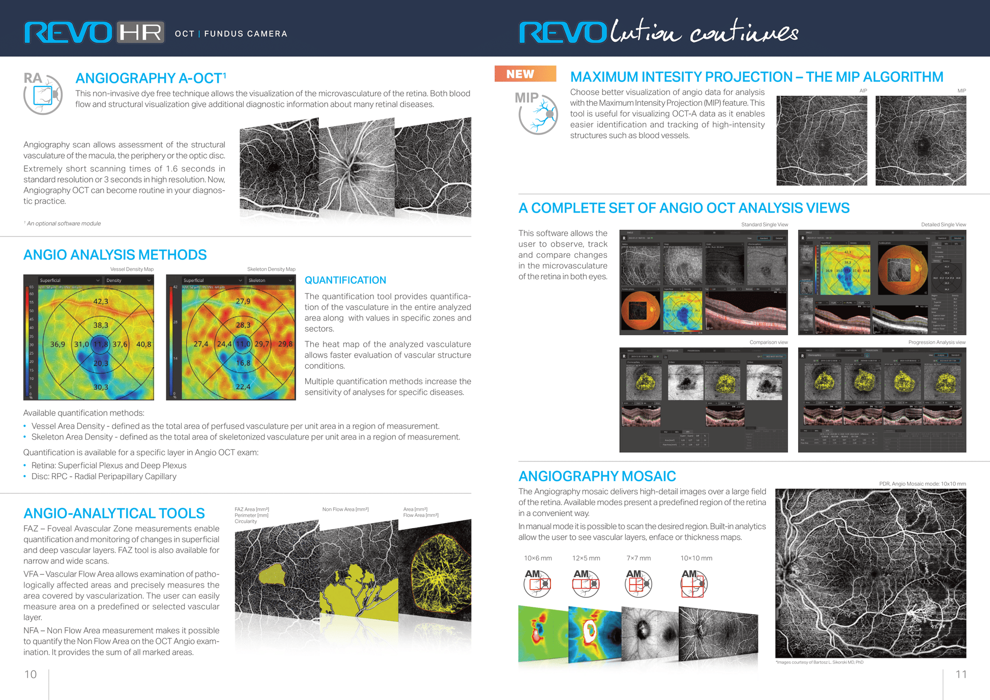
The REVO HR is an All in One device you can use in a number of ways such as a full color Fundus Camera or as a combo,
providing simultaneous OCT and fundus images for high quality OCT imaging, including OCT-A.
multiple Functions in One device
OCT made simple as never beFOre
Simply position the patient and press the START button to
acquire examinations of both eyes. The REVO HR guides the
patient through the process with voice commands, which
increases comfort and reduces patient chair time.
a perFeCT FiT FOr every praCTiCe
With its small system footprint and single cable connection, the
REVO HR can be placed in the smallest of exam rooms. The
REVO can easily function as both a screening or an advanced di-
agnostic device with its variety of examination and analysis tools.AccuTrack TM
Real time hardware eye tracking
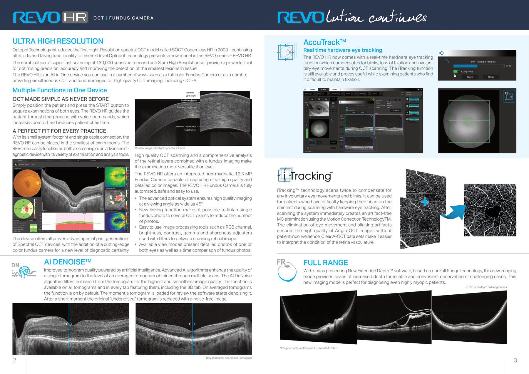
The REVO HR now comes with a real-time hardware eye tracking
function which compensates for blinks, loss of fixation and involun-
tary eye movements during OCT scanning. The iTracking function
is still available and proves useful while examining patients who find
it difficult to maintain fixation.
iTracking™ technology scans twice to compensate for
any involuntary eye movements and blinks. It can be used
for patients who have difficulty keeping their head on the
chinrest during scanning with hardware eye tracking. After,
scanning the system immediately creates an artifact-free
MC examination using the Motion Correction TechnologyTM.
The elimination of eye movement and blinking artifacts
ensures the high quality of Angio OCT images without
patient inconvenience. Clear A-OCT data sets make it easier
to interpret the condition of the retina vasculature.
Raw Tomogram / AiDenoise Tomogram
ai denOiseTm
Improved tomogram quality powered by artificial intelligence. Advanced AI algorithms enhance the quality of
a single tomogram to the level of an averaged tomogram obtained through multiple scans. The AI DeNoise
algorithm filters out noise from the tomogram for the highest and smoothest image quality. The function is
available on all tomograms and in every tab featuring them, including the 3D tab. On averaged tomograms
the function is on by default. The moment a tomogram is loaded for review the software starts denoising it.
After a short moment the original “undenoised” tomogram is replaced with a noise-free image.
*Images courtesy of Bartosz L. Sikorski MD, PhD
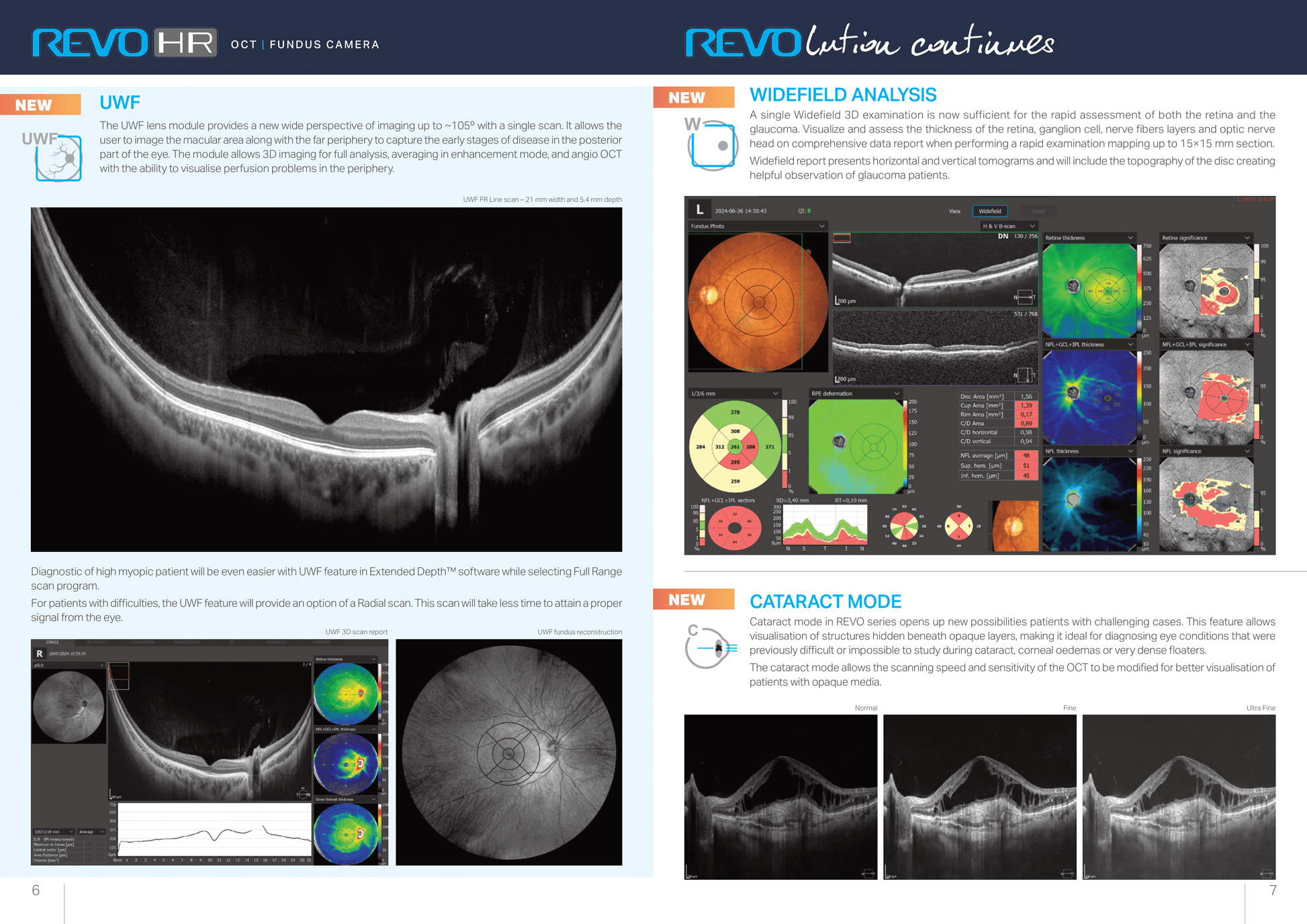
Full ranGe
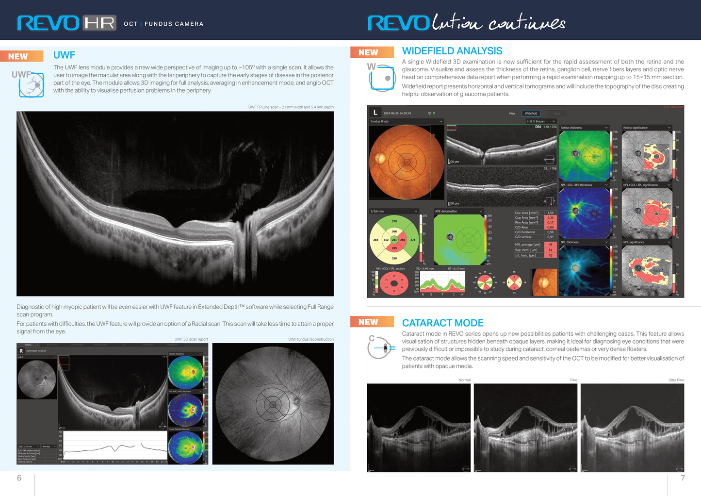
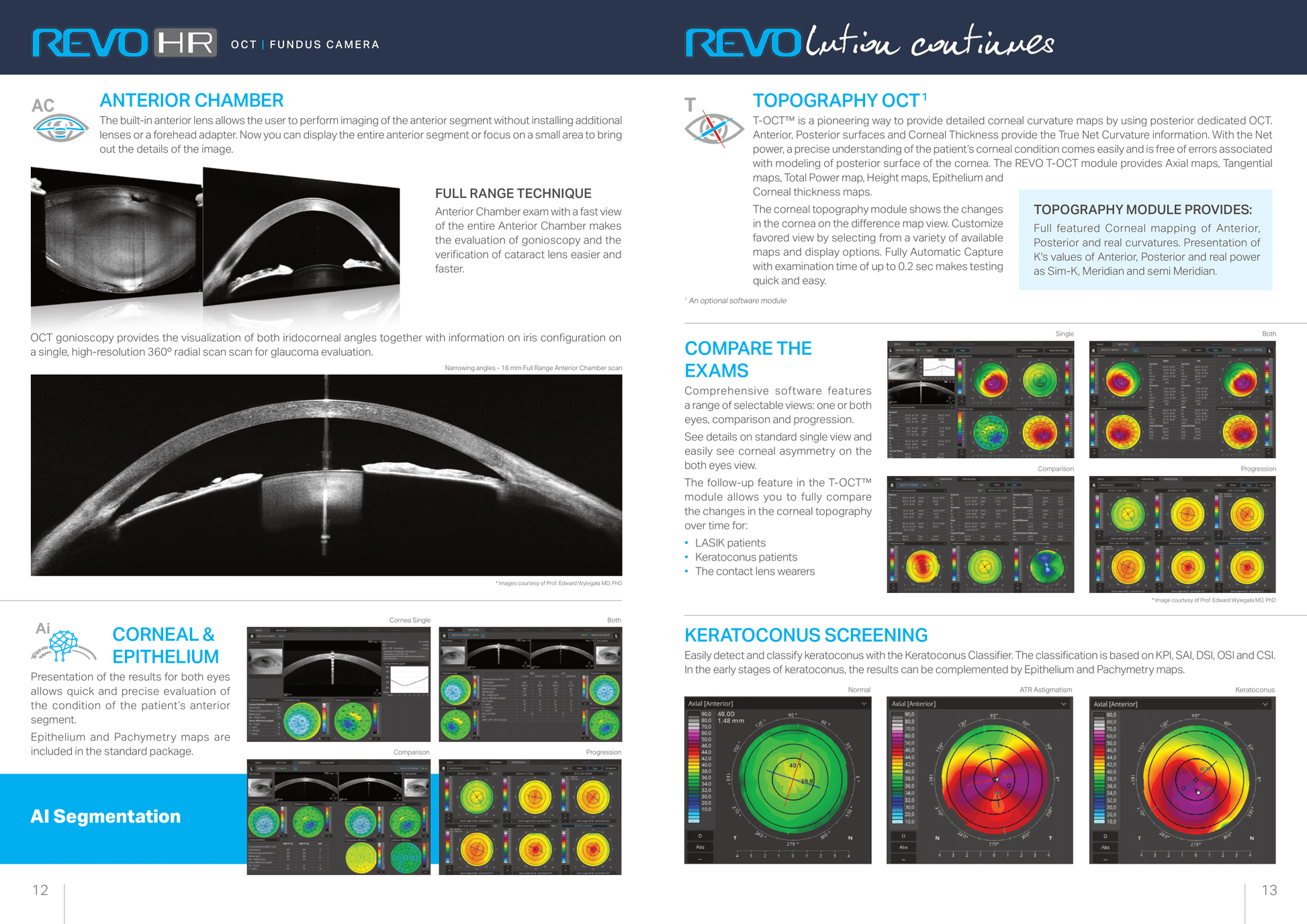
With scans presenting New Extended Depth™ software, based on our Full Range technology, this new imaging
mode provides scans of increased depth for reliable and convenient observation of challenging cases. This
new imaging mode is perfect for diagnosing even highly myopic patients.
Anterior line 9 mm
Tear film
Epithelium
Bowman’s membrane
Stroma
Descemet’s membrane
Endothelium
Basal membrane

  • Easy Setup
  • 24/7 VIP Support
View full details

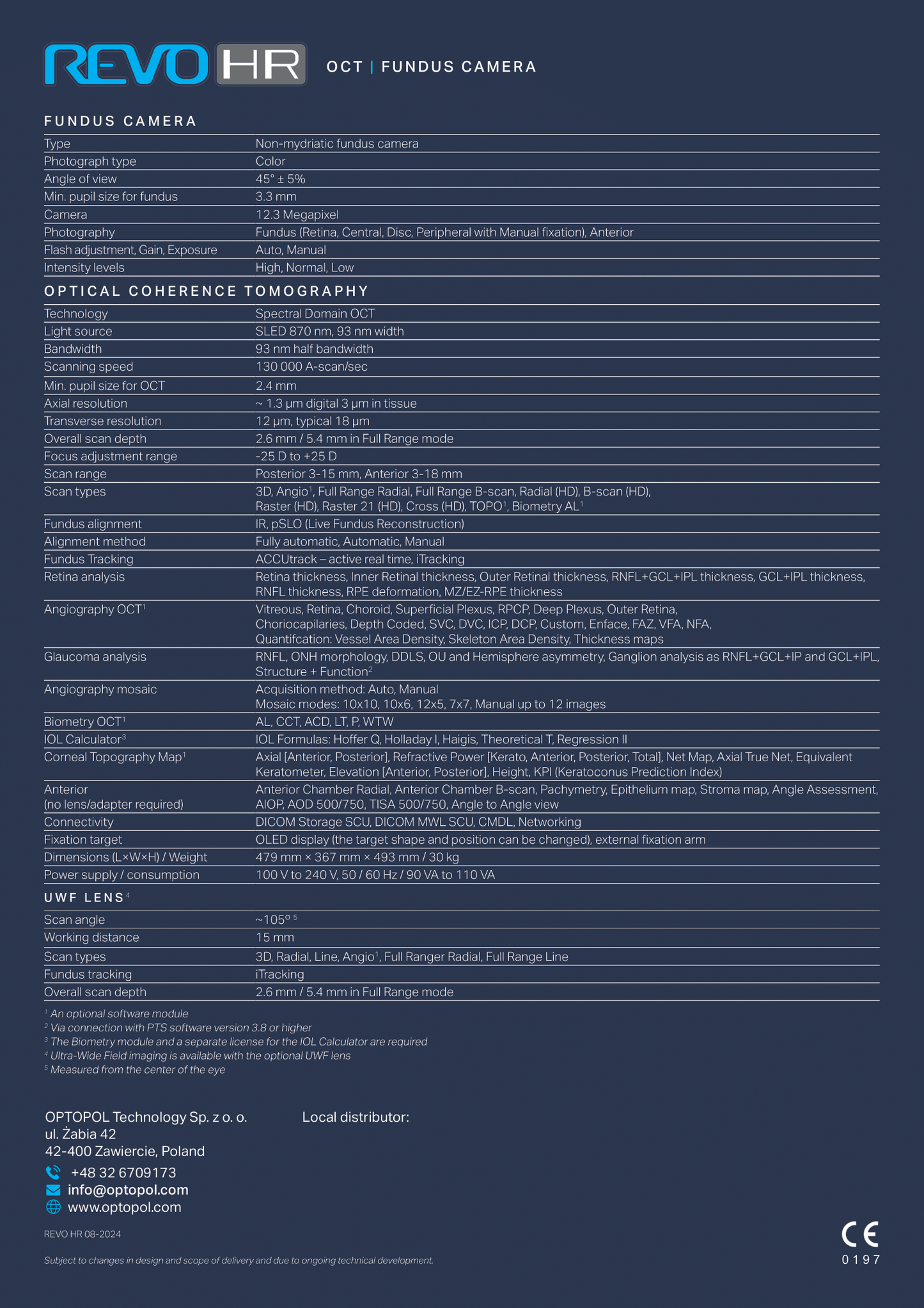
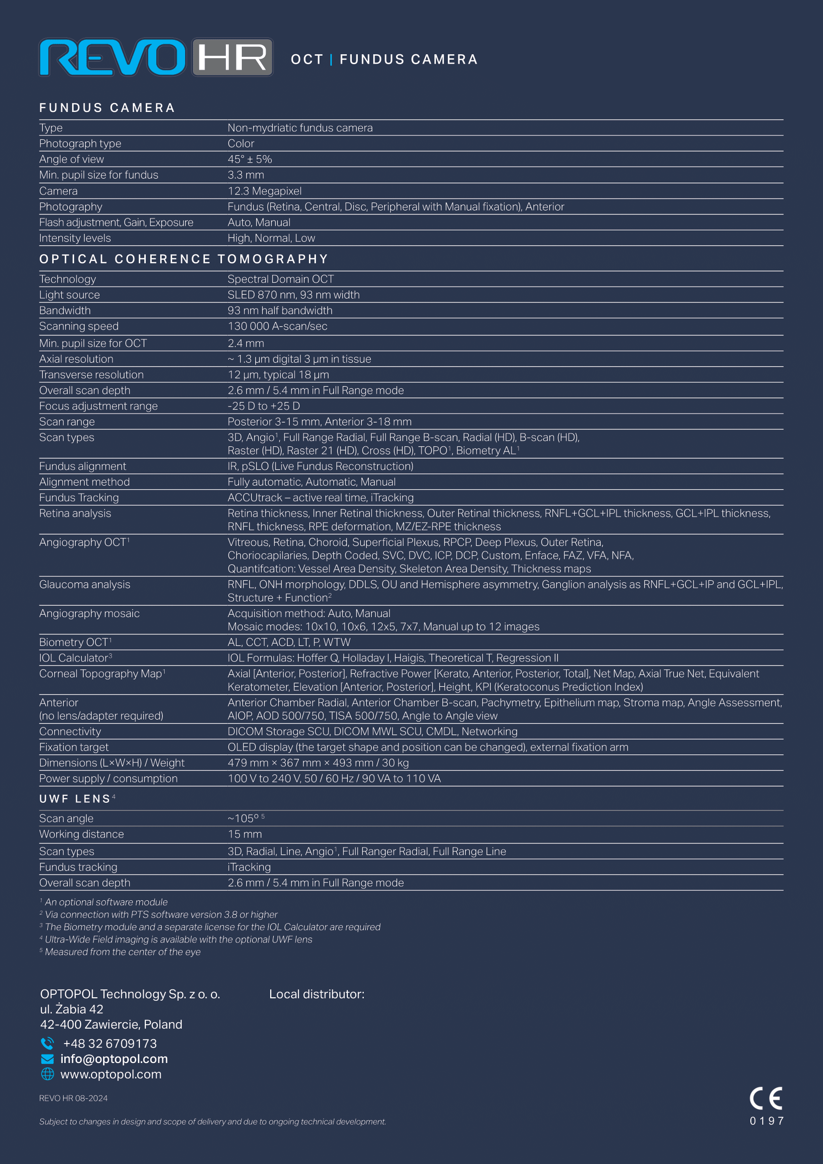
Key Specifications

Scanning Speed

130,000 A-scans per second

Resolution

3 µm optical resolution

Fundus Camera

12.3 MP non-mydriatic color imaging

Eye Tracking

AccuTrack™ real-time hardware eye tracking

AI Features

AI-DENOISE™ image enhancement

Scan Capabilities

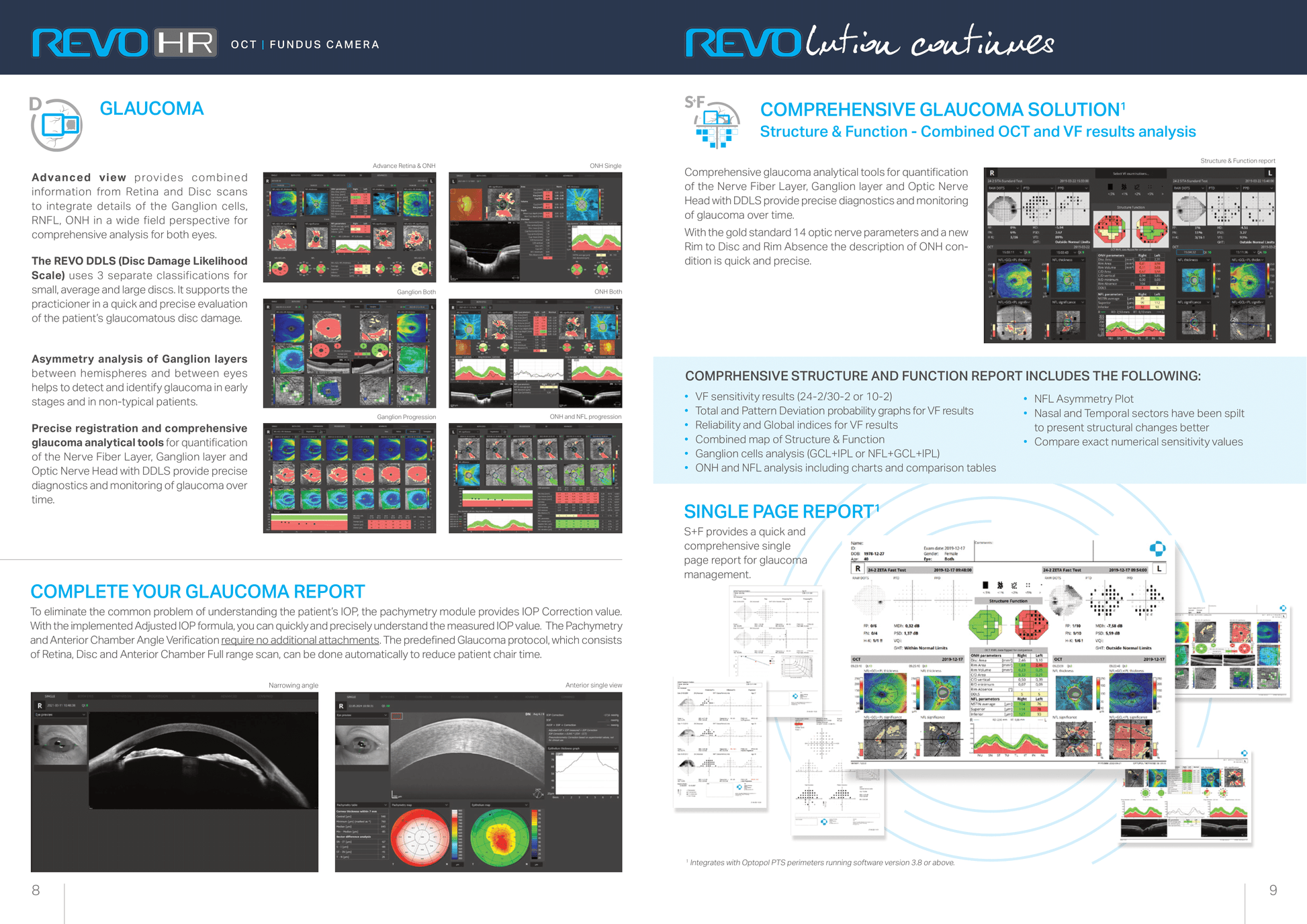
Posterior Segment: Retina, optic nerve head, macula

Anterior Segment: Cornea, epithelium, pachymetry

Optional Modules

OCT-Angiography (OCT-A)

T-OCT™ Topography

B-OCT® Biometry

IOL Calculations

Ultra-Widefield (UWF) Imaging

User Interface

Fully automated operation with single-button capture

Connectivity

DICOM, USB, and network compatibility

Physical Dimensions

Compact design suitable for small exam rooms

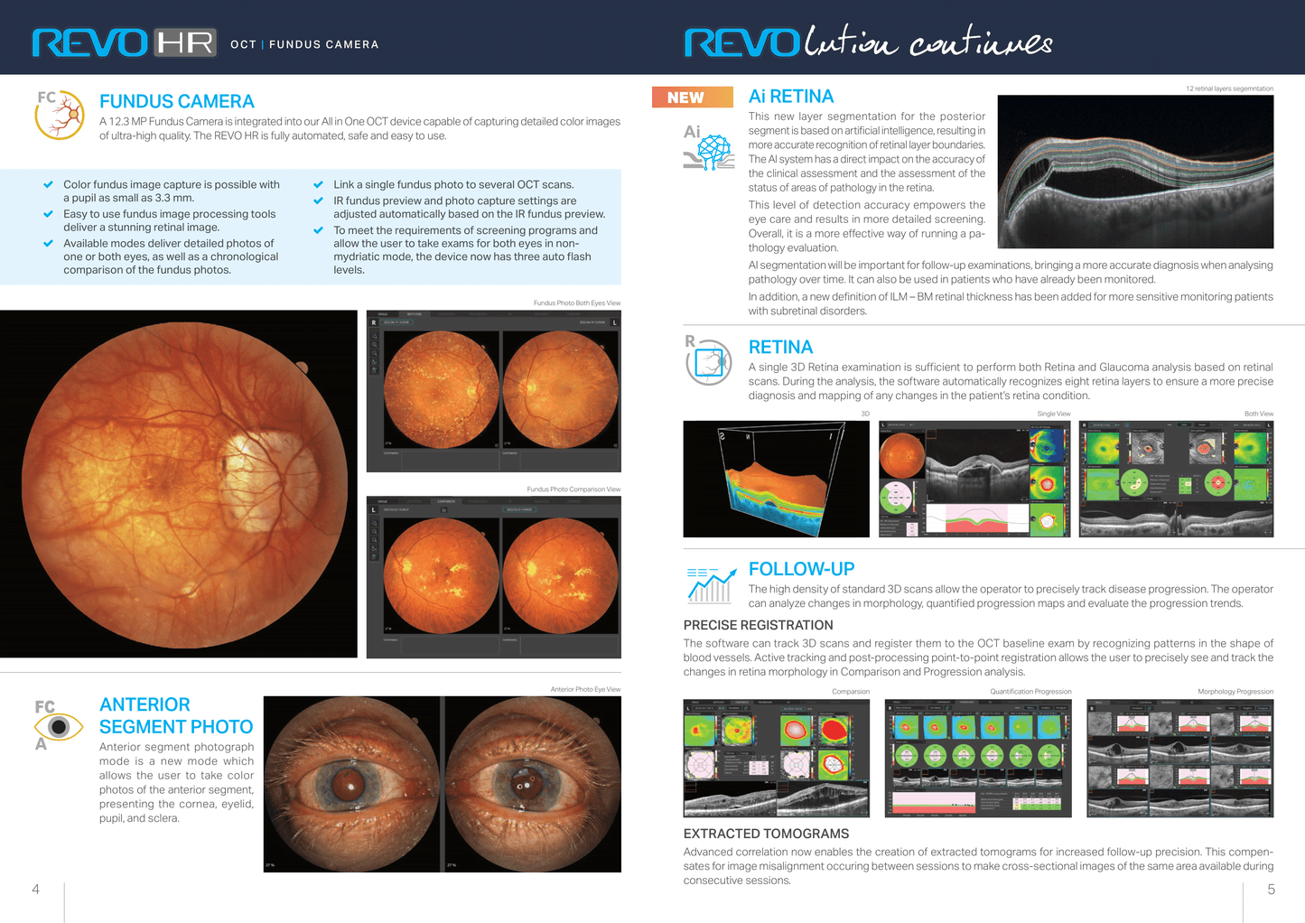
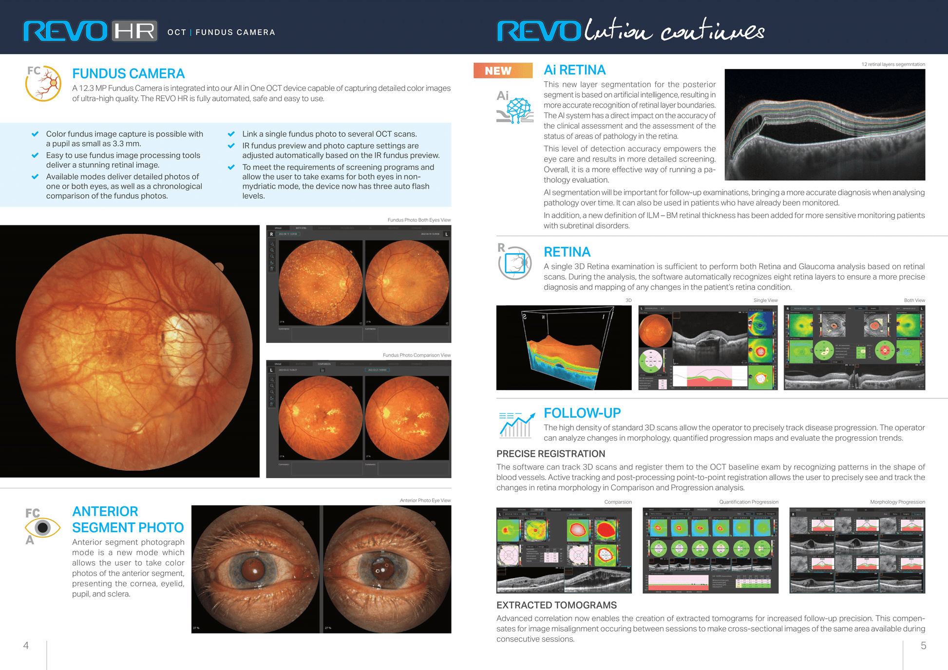
Fundus Camera & AI Retina Technology

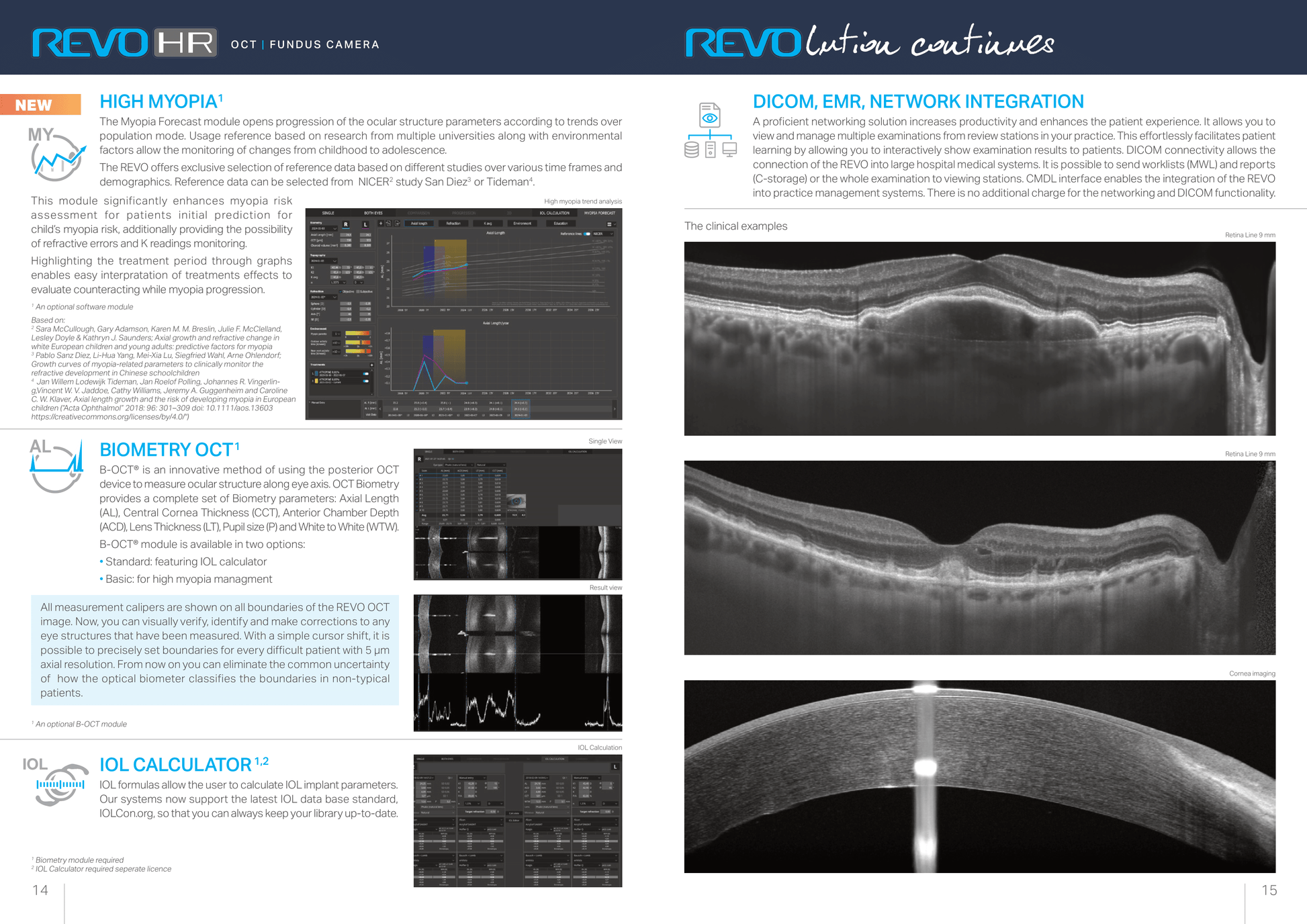
The REVO HR features a 12.3 MP non-mydriatic color fundus camera that delivers ultra-clear, true-to-life retinal images without the need for pupil dilation, ensuring faster and more comfortable exams. It captures wide-field views of the macula, optic nerve head, and peripheral retina with sharp detail—essential for accurate diagnosis and monitoring.

Paired with powerful AI Retina technology, the device automatically detects, highlights, and quantifies retinal abnormalities like microaneurysms, hemorrhages, and exudates. This AI-driven analysis accelerates diagnosis, supports early disease detection, and enhances treatment planning by providing consistent, reliable insights right at the point of care.

AccuTrack, AI-DENOISE & Dual Range UHD Technology

The REVO HR is engineered with AccuTrack™ real-time hardware eye tracking, a cutting-edge system that continuously monitors and compensates for even the slightest eye movements during scanning. This real-time tracking significantly reduces motion artifacts and blurring, which are common challenges in retinal imaging. By maintaining perfect alignment between the scanning beam and the retina, AccuTrack™ ensures that every image captured is crisp, stable, and clinically reliable, even in patients who find it hard to fixate steadily. This level of precision is vital for accurate diagnosis and longitudinal monitoring, especially when tracking subtle changes over time in chronic eye conditions like glaucoma or diabetic retinopathy.

Complementing the hardware tracking is AI-DENOISE™, an advanced artificial intelligence-powered noise reduction technology that enhances image quality by intelligently filtering out background noise and artifacts without sacrificing critical anatomical details. Unlike traditional noise reduction methods that can blur or distort images, AI-DENOISE™ uses deep learning algorithms trained on vast datasets of retinal images to differentiate between true signal and noise. The result is ultra-clear, high-contrast OCT scans that reveal microstructures of the retina with exceptional clarity. This enhancement empowers clinicians to detect subtle pathologies earlier and with greater confidence, improving patient outcomes through timely intervention.

The Dual Range UHD (Ultra High Definition) imaging capability further elevates the REVO HR’s diagnostic power by combining the best of both worlds: wide-field scanning for comprehensive retinal overview and ultra-high-resolution scans for detailed analysis of specific regions. This dual-mode approach means that clinicians no longer have to choose between coverage and detail—they get both in one seamless workflow. The wide-field scans provide context and help identify areas of concern across the macula, optic nerve head, and peripheral retina, while the UHD scans zoom in to highlight microstructural changes and fine abnormalities with stunning precision. This versatility supports a broad spectrum of ophthalmic assessments, from routine screenings to complex retinal disease evaluations.

Together, these technologies make the REVO HR a powerhouse device that delivers unparalleled image fidelity, diagnostic accuracy, and clinical confidence—allowing eye care professionals to provide faster, more precise, and personalized care with every patient.